
Page 72 - Genetics_From_Genes_to_Genomes_6th_FULL_Part2
P. 72
7.2 Molecular Mechanisms That Alter DNA Sequence 231
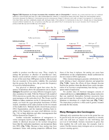
Figure 7.13 Exposure to X-rays increases the mutation rate in Drosophila. F 1 females are constructed that have an irradiated
paternal X chromosome (red line) and a Bar-marked Balancer maternal X chromosome (wavy blue line). These two chromosomes cannot
recombine because the Balancer chromosome prevents crossing-over. Single F 1 females, each with a single X-ray-exposed X chromosome
from their father, are then individually mated with wild-type males. If the paternal X chromosome in any one F 1 female has an X-ray-induced
recessive lethal mutation (m), she can produce only Bar-eye sons (left). If the X chromosome has no such mutation, this F 1 female will
produce both Bar-eye and non-Bar-eye sons (right).
X-rays
Bar
P X X
Y X
X
F Bar-eye females Bar
1
X
Individual matings:
F 1 Bar-eye Wild type F Bar-eye Wild type
1
Bar x Bar x
If F female has an X-ray- If F female has no X-ray-
1
1
induced recessive lethal on X induced recessive lethal on X
m Bar Bar
(Dies) Bar-eye Non-Bar-eye Bar-eye
unable to produce non-Bar-eye sons. Thus, simply by those of the base it replaces, the analog can cause base
noting the presence or absence of non-Bar-eye sons, substitutions on the complementary strand synthesized in
Muller could establish whether a mutation had occurred the next round of DNA replication.
in any of the more than 1000 genes on the X chromosome Other chemical mutagens generate substitutions by di-
that are essential to Drosophila viability. He concluded rectly altering a base’s chemical structure and properties
that the greater the X-ray dose, the greater the frequency (Fig. 7.14b). Again, the effects of these changes become
of recessive lethal mutations. fixed in the genome when the altered base causes incorpo-
Any physical or chemical agent that raises the fre- ration of an incorrect complementary base during a subse-
quency of mutations above the spontaneous rate is called a quent round of replication.
mutagen. Researchers use many different mutagens to pro- Yet another class of chemical mutagens consists of
duce mutations for study. With the Watson-Crick model of compounds known as intercalators: flat, planar molecules
DNA structure as a guide, they can understand the action of that can sandwich themselves between successive base
most mutagens at the molecular level. The X-rays used by pairs and disrupt the machinery for replication, generating
Muller to induce mutations on the X chromosome, for ex- deletions or insertions of a single base pair (Fig. 7.14c).
ample, can break the sugar-phosphate backbones of DNA The intercalator proflavin is often used in genetic research
strands, sometimes at the same position on the two strands precisely for this reason.
of the double helix. Multiple double-strand breaks produce
DNA fragmentation, and the improper stitching back to-
gether of the fragments can cause small deletions (review Many Mutagens Are Carcinogens
Fig. 7.8c) or large deletions and other rearrangements that
will be discussed in Chapter 13. Although only mutations that occur in the germ line can
Another molecular mechanism of mutagenesis in- be passed on to the next generation, mutations in somatic
volves mutagens known as base analogs, which are so cells can still have an impact on the well-being and
similar in chemical structure to the normal nitrogenous survival of individuals. Somatic mutations in genes that
bases that the replication machinery can incorporate them help regulate the cell cycle may, for example, lead to
into DNA (Fig. 7.14a). Because a base analog may have cancer. For this reason, many mutagens act as carcinogens
tautomeric forms with pairing properties different from (cancer-causing agents).