
Page 77 - Genetics_From_Genes_to_Genomes_6th_FULL_Part2
P. 77
236 Chapter 7 Anatomy and Function of a Gene: Dissection Through Mutation
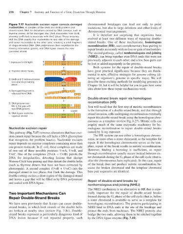
Figure 7.17 Nucleotide excision repair corrects damaged chromosomal breakages can lead not only to point
nucleotides. A complex of the UvrA and UvrB proteins (not mutations, but also to large deletions and other kinds of
shown) scans DNA for distortions caused by DNA damage, such as chromosomal rearrangements.
thymine dimers. At the damaged site, UvrA dissociates from UvrB, It is therefore not surprising that organisms have
allowing UvrB (red) to associate with UvrC (blue). These enzymes
nick the DNA exactly four nucleotides to one side of the damage evolved at least two different ways of repairing double-
and seven nucleotides to the other side, releasing a small fragment strand breaks. One of these mechanisms, homologous
of single-stranded DNA. DNA polymerases then resynthesize the recombination (HR), uses complementary base pairing to
missing information (green), and DNA ligase reseals the now- repair breaks accurately with no loss or gain of nucleotides.
corrected strand. The second pathway, called nonhomologous end-joining
UV
(NHEJ), can bring together even DNA ends that were not
previously adjacent to each other, and a few base pairs can
1. Exposure to UV light 5' T T 3' be lost or added improperly in the process.
3' AA 5'
Both systems for the repair of double-strand breaks
2. Thymine dimer forms. T T have great practical significance because they are funda-
AA mental to new, effective strategies for genome editing (al-
3. UvrB and C endonucleases Nick Nick tering an organism’s genome in specific ways). We will
nick strand containing T T describe these exciting methods for modifying genomes in
dimer. AA Chapter 18, but it will be helpful for you to gain here some
idea about how these repair mechanisms work.
4. Damaged fragment is T T
released from DNA.
AA Double-strand break repair via homologous
recombination (HR)
5. DNA polymerase
fills in the gap with TT You will recall that the first step of meiotic recombination
new DNA (green ). AA
Ligase is the formation of a double-strand break, and that through
6. DNA ligase seals the strand invasion, cells undergoing recombination eventually
repaired strand. TT
AA repair this double-strand break using the homologous chro-
mosome as a template (review Fig. 6.27). Mitotic cells can
employ much of the same enzymatic machinery for ho-
Nucleotide excision repair mologous recombination to repair double-strand breaks
This pathway (Fig. 7.17) removes alterations that base exci- caused by X-ray exposure.
sion cannot repair because the cell lacks a DNA glycosylase The HR system can use either a homologous chromo-
that recognizes the problem base(s). Nucleotide excision some, or more often a sister chromatid, as the template for
repair depends on enzyme complexes containing more than repair. If the homologous chromosome serves as the tem-
one protein molecule. In E. coli, these complexes are made plate, repair of the break results in mitotic recombination.
of two out of three possible proteins: UvrA, UvrB, and However, finding a homolog is inefficient, so repair
UvrC. One of the complexes (UvrA + UvrB) patrols the through recombination usually occurs instead between sis-
DNA for irregularities, detecting lesions that disrupt ter chromatids during the G 2 phase of the cell cycle (that is,
Watson-Crick base pairing and thus distort the double helix after the chromosomes have replicated). In this case, repair
(such as thymine dimers that have not been corrected by of the break does not produce mitotic recombination be-
photorepair). A second complex (UvrB + UvrC) cuts the cause the broken chromatid and the template chromatid
damaged strand in two places that flank the damage. This base pair sequences are identical.
double-cutting excises a short region of the damaged strand
and leaves a gap that will be filled in by DNA polymerase Repair of double-strand breaks by
and sealed with DNA ligase.
nonhomologous end-joining (NHEJ)
The NHEJ mechanism is an alternative to HR that is espe-
Two Important Mechanisms Can cially important for the repair of double-strand breaks
Repair Double-Strand Breaks formed during the G 1 phase of the cell cycle (that is, before
a sister chromatid is available to serve as a template for
We have seen previously that X-rays can cause double- homologous recombination). The proteins participating in
strand breaks, in which both strands of the double helix NHEJ bind to DNA ends at the site of the breakage and
are broken at nearby sites (review Fig. 7.8c). Double- protect the ends from nucleases. The NHEJ proteins also
strand breaks represent a particularly dangerous kind of bridge the two ends, allowing them to be stitched together
DNA lesion because if not repaired properly, such by the DNA ligase enzyme (Fig. 7.18).