
Page 81 - Genetics_From_Genes_to_Genomes_6th_FULL_Part3
P. 81
11.2 Genotyping a Known Disease-Causing Mutation 375
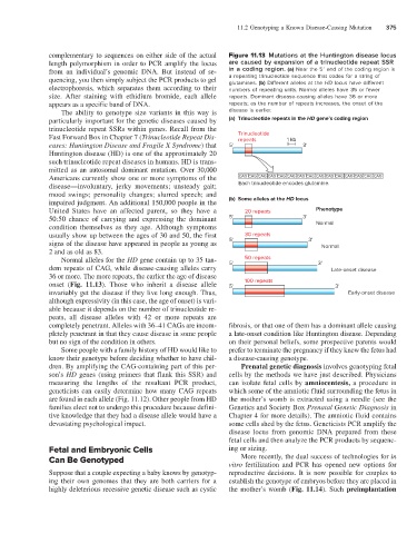
complementary to sequences on either side of the actual Figure 11.13 Mutations at the Huntington disease locus
length polymorphism in order to PCR amplify the locus are caused by expansion of a trinucleotide repeat SSR
from an individual’s genomic DNA. But instead of se- in a coding region. (a) Near the 5′ end of the coding region is
quencing, you then simply subject the PCR products to gel a repeating trinucleotide sequence that codes for a string of
glutamines. (b) Different alleles at the HD locus have different
electrophoresis, which separates them according to their numbers of repeating units. Normal alleles have 35 or fewer
size. After staining with ethidium bromide, each allele repeats. Dominant disease-causing alleles have 36 or more
appears as a specific band of DNA. repeats; as the number of repeats increases, the onset of the
The ability to genotype size variants in this way is disease is earlier.
particularly important for the genetic diseases caused by (a) Trinucleotide repeats in the HD gene’s coding region
trinucleotide repeat SSRs within genes. Recall from the
Fast Forward Box in Chapter 7 (Trinucleotide Repeat Dis- Trinucleotide 1 kb
repeats
eases: Huntington Disease and Fragile X Syndrome) that 5' 3'
Huntington disease (HD) is one of the approximately 20
such trinucleotide repeat diseases in humans. HD is trans-
mitted as an autosomal dominant mutation. Over 30,000
Americans currently show one or more symptoms of the CAG CAG CAG CAG CAG CAG CAG CAG CAG CAG CAG CAG CAG CAG CAG
disease—involuntary, jerky movements; unsteady gait; Each trinucleotide encodes glutamine.
mood swings; personality changes; slurred speech; and
impaired judgment. An additional 150,000 people in the (b) Some alleles at the HD locus
United States have an affected parent, so they have a 20 repeats Phenotype
50:50 chance of carrying and expressing the dominant 5' 3'
condition themselves as they age. Although symptoms Normal
usually show up between the ages of 30 and 50, the first 30 repeats
signs of the disease have appeared in people as young as 5' 3' Normal
2 and as old as 83.
Normal alleles for the HD gene contain up to 35 tan- 50 repeats
dem repeats of CAG, while disease-causing alleles carry 5' 3' Late-onset disease
36 or more. The more repeats, the earlier the age of disease
onset (Fig. 11.13). Those who inherit a disease allele 5' 100 repeats 3'
invariably get the disease if they live long enough. Thus, Early-onset disease
although expressivity (in this case, the age of onset) is vari-
able because it depends on the number of trinucleotide re-
peats, all disease alleles with 42 or more repeats are
completely penetrant. Alleles with 36–41 CAGs are incom- fibrosis, or that one of them has a dominant allele causing
pletely penetrant in that they cause disease in some people a late-onset condition like Huntington disease. Depending
but no sign of the condition in others. on their personal beliefs, some prospective parents would
Some people with a family history of HD would like to prefer to terminate the pregnancy if they knew the fetus had
know their genotype before deciding whether to have chil- a disease-causing genotype.
dren. By amplifying the CAG-containing part of this per- Prenatal genetic diagnosis involves genotyping fetal
son’s HD genes (using primers that flank this SSR) and cells by the methods we have just described. Physicians
measuring the lengths of the resultant PCR product, can isolate fetal cells by amniocentesis, a procedure in
geneticists can easily determine how many CAG repeats which some of the amniotic fluid surrounding the fetus in
are found in each allele (Fig. 11.12). Other people from HD the mother’s womb is extracted using a needle (see the
families elect not to undergo this procedure because defini- Genetics and Society Box Prenatal Genetic Diagnosis in
tive knowledge that they had a disease allele would have a Chapter 4 for more details). The amniotic fluid contains
devastating psychological impact. some cells shed by the fetus. Geneticists PCR amplify the
disease locus from genomic DNA prepared from these
fetal cells and then analyze the PCR products by sequenc-
Fetal and Embryonic Cells ing or sizing.
Can Be Genotyped More recently, the dual success of technologies for in
vitro fertilization and PCR has opened new options for
Suppose that a couple expecting a baby knows by genotyp- reproductive decisions. It is now possible for couples to
ing their own genomes that they are both carriers for a establish the genotype of embryos before they are placed in
highly deleterious recessive genetic disease such as cystic the mother’s womb (Fig. 11.14). Such preimplantation