
Page 82 - Genetics_From_Genes_to_Genomes_6th_FULL_Part3
P. 82
376 Chapter 11 Analyzing Genomic Variation
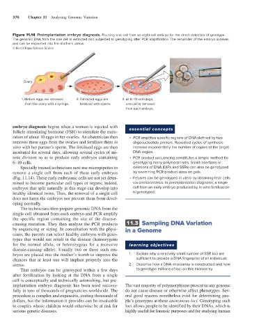
Figure 11.14 Preimplantation embryo diagnosis. Plucking one cell from an eight-cell embryo for the direct detection of genotype.
The genomic DNA from the one cell is extracted and subjected to genotyping after PCR amplification. The remainder of the embryo survives
and can be implanted into the mother’s uterus.
© Benoît Rajau/Science Source
Ovary
1. Mature eggs are retrieved 2. Extracted eggs are 3. At 6–10-cell stage,
from the ovary with a syringe. fertilized with sperm. one cell is removed
from each embryo.
embryo diagnosis begins when a woman is injected with essential concepts
follicle stimulating hormone (FSH) to stimulate the matu-
ration of about 10 eggs in her ovaries. An obstetrician then • PCR amplifies specific regions of DNA defined by two
removes these eggs from the ovaries and fertilizes them in oligonucleotide primers. Repeated cycles of synthesis
vitro with her partner’s sperm. The fertilized eggs are then increase exponentially the number of copies of the target
incubated for several days, allowing several cycles of mi- DNA region.
totic division so as to produce early embryos containing • PCR product sequencing constitutes a simple method for
6–10 cells. genotyping many polymorphisms. Small insertions or
Specially trained technicians next use micropipettes to deletions of DNA (DIPs and SSRs) can also be genotyped
remove a single cell from each of these early embryos by examining PCR product sizes on gels.
(Fig. 11.14). These early embryonic cells are not yet deter- • Fetuses can be genotyped in utero by obtaining fetal cells
mined to become particular cell types or organs; indeed, via amniocentesis. In preimplantation diagnosis, a single
embryos that split naturally at this stage can develop into cell from an early embryo produced by in vitro fertilization
healthy identical twins. Thus, the removal of a single cell is genotyped.
does not harm the embryos nor prevent them from devel-
oping normally.
The technicians then prepare genomic DNA from the
single cell obtained from each embryo and PCR amplify
the specific region containing the site of the disease-
causing mutation. They then analyze the PCR products 11.3 Sampling DNA Variation
by sequencing or sizing. In consultation with the physi- in a Genome
cians, the parents can select healthy embryos with geno-
types that would not result in the disease (homozygous
for the normal allele, or heterozygous for a recessive learning objectives
disease-causing allele). Usually two or three such em-
bryos are placed into the mother’s womb to improve the 1. Explain why a relatively small number of SSR loci are
chances that at least one will implant properly into the sufficient to provide a DNA fingerprint of an individual.
uterus. 2. Describe how a DNA microarray is constructed and how
That embryos can be genotyped within a few days to genotype millions of loci on this microarray.
after fertilization by looking at the DNA from a single
cell is conceptually and technically astonishing, but pre-
implantation embryo diagnosis has been used success- The vast majority of polymorphisms present in any genome
fully in tens of thousands of pregnancies worldwide. The do not cause disease or otherwise affect phenotypes. Sev-
procedure is complex and expensive, costing thousands of eral good reasons nonetheless exist for determining peo-
dollars, but the information it provides can be invaluable ple’s genotypes at these anonymous loci. Genotyping such
to couples whose children would otherwise be at risk for loci allows people to be identified by their DNAs, which is
serious genetic diseases. highly useful for forensic purposes and for studying human