
Page 97 - Genetics_From_Genes_to_Genomes_6th_FULL_Part2
P. 97
256 Chapter 7 Anatomy and Function of a Gene: Dissection Through Mutation
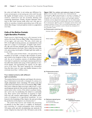
the color red looks like, to not seeing any difference be- Figure 7.32 The cellular and molecular basis of vision.
tween red and green, to not seeing any color at all. Second, (a) Rod and cone cells in the retina carry membrane-bound
the highly developed science of psychophysics provides photoreceptors. (b) The photoreceptor in rod cells is rhodopsin. The
sensitive, noninvasive tests for accurately defining and blue, green, and red receptor proteins in cone cells are related to
rhodopsin. The colored dots are amino acids that differ between
comparing phenotypes. Finally, because inherited varia- rhodopsin and the diagrammed protein. (c) One red photoreceptor
tions in the visual system rarely affect one’s life span or gene and one to three green photoreceptor genes are clustered on
ability to reproduce, mutations generating many of the new the X chromosome. (d) The genes for rhodopsin and the three color
alleles that change visual perception remain in a population receptors probably evolved from a primordial photoreceptor gene
over time. through three gene duplication events followed by divergence of
the duplicated copies.
(a) Photoreceptor-containing cells
Rod and cone cells Pigmented
Cells of the Retina Contain epithelium
Light-Sensitive Proteins
People perceive light through nerve cells (neurons) in the
retina at the back of the eye (Fig. 7.32a). These neurons are Retina surface
of two types: rods and cones. The rods, which make up
95% of all light-receiving neurons, are stimulated by weak
light over a range of wavelengths. At higher light intensi- Light
ties, the rods become saturated and no longer send mean- Photoreceptor
cells
ingful information to the brain. This is when the cones take Rod
over, processing wavelengths of bright light that enable us
to see color.
The cones come in three forms—one specializes in the
reception of red light, a second in the reception of green, Cone
and a third in the reception of blue. For each photoreceptor Disc membrane
cell, the act of reception consists of absorbing photons
from light of a particular wavelength, transducing informa- Light
tion about the number and energy of those photons to elec- Retinal
trical signals, and transmitting the signals via the optic
nerve to the brain. The brain integrates the information Rhodopsin Membranous disc
from the three types of cones and enables humans to dis-
criminate more than 1 million colors.
(b) Photoreceptor proteins
Rhodopsin protein Blue-receiving protein
Four related proteins with different C C
light sensitivities
The protein that receives photons and triggers the process-
ing of information in rod cells is rhodopsin. It consists of a
single polypeptide chain containing 348 amino acids that N N
snakes back and forth across the cell membrane (Fig. 7.32b). Green-receiving protein Red-receiving protein
C
C
One lysine within the chain associates with retinal, a carot-
enoid pigment molecule that actually absorbs photons. The
amino acids in the vicinity of the retinal constitute rhodop-
sin’s active site; by positioning the retinal in a particular
way, those amino acids determine its response to light. N N
Each rod cell contains approximately 100 million mole-
cules of rhodopsin in its specialized membrane. As you
learned at the beginning of this chapter, the gene governing (c) Red/green pigment genes (d) Evolution of
the production of rhodopsin is on chromosome 3. X chromosomes visual pigment genes
The protein that receives and initiates the processing from normal individuals: Primordial gene
of photons in the blue cones is a relative of rhodopsin,
also consisting of a single polypeptide chain containing
348 amino acids and also encompassing one molecule of
retinal. Slightly less than half of the 348 amino acids in the
Red Green Blue Rhodopsin
gene gene gene gene