
Page 99 - Genetics_From_Genes_to_Genomes_6th_FULL_Part2
P. 99
258 Chapter 7 Anatomy and Function of a Gene: Dissection Through Mutation
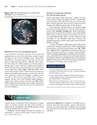
Figure 7.34 How the world looks to a person with Unequal crossing-over between
tritanopia. Compare with Fig. 4.22. the red and green genes
Color deficit simulation courtesy of Vischeck (www.vischeck.com). Source image
courtesy of NASA People with normal color vision have a single red gene;
some of these normal individuals also have a single adja-
cent green gene, while others have two or even three green
genes. The red and green genes are 96% identical in DNA
sequence; the different green genes, 99.9% identical.
Their proximity and high degree of homology make
these genes unusually prone to an error in meiotic recombi-
nation called unequal crossing-over. When homologous
chromosomes associate during meiosis, two closely related
DNA sequences that are adjacent to each other, like the red
and green photoreceptor genes, can pair with each other
incorrectly. If recombination takes place between the
mispaired sequences, photoreceptor genes may be deleted,
added, or changed.
A variety of unequal recombination events produce DNA
containing no red gene, no green gene, various combinations
of green genes, or hybrid red-green genes (see Fig. 7.33d).
These different DNA combinations account for the large ma-
Mutations in the cone cell pigment genes jority of the known aberrations in red-green color perception,
Vision problems caused by mutations in the cone cell with the remaining abnormalities stemming from point muta-
pigment genes are less severe than those caused by simi- tions, as described earlier. Because the accurate perception of
lar defects in the rod cell rhodopsin gene. Most likely, red and green depends on the differing ratios of red and green
this difference occurs because the rods make up 95% of a light processed, people with no red or no green gene perceive
person’s light-receiving neurons, while the cones consti- red and green as the same color (see Fig. 4.22).
tute only about 5%. Some mutations in the blue gene on
chromosome 7 cause tritanopia, a defect in the ability to essential concepts
discriminate between colors that differ only in the amount
of blue light they contain (Figs. 7.33b and 7.34). Muta- • The vision pigments in humans consist of the protein
tions in the red gene on the X chromosome can modify or rhodopsin in rods plus the blue-, red-, and green-sensitive
abolish red protein function and as a result, the red cone photoreceptors in cones.
cells’ sensitivity to light. For example, a change at posi- • The four genes of the rhodopsin family evolved from an
tion 203 in the red-receiving protein from cysteine to ancestral photoreceptor gene by successive rounds of
arginine disrupts one of the disulfide bonds required to gene duplication and divergence.
support the protein’s tertiary structure (see Fig. 7.33c). • Mutations in the rhodopsin gene may disrupt rod function,
Without that bond, the protein cannot stably maintain its leading to blindness. Mutations in cone cell photoreceptor
native configuration, and a person with the mutation has genes are responsible for various forms of color blindness.
red color blindness.
WHAT’S NEXT
Careful studies of mutations showed that genes are linear polypeptide colinearity. In Chapter 8, we explain how co-
arrays of mutable elements that direct the assembly of linearity arises from base pairing, a genetic code, specific
amino acids in a polypeptide. The mutable elements are the enzymes, and macromolecular assemblies like ribosomes
nucleotide building blocks of DNA. that guide the flow of information from DNA through RNA
Biologists call the parallel between the sequence of to protein.
nucleotides in a gene and the order of amino acids in a
DNA: © Design Pics/Bilderbuch RF