
Page 94 - Genetics_From_Genes_to_Genomes_6th_FULL_Part2
P. 94
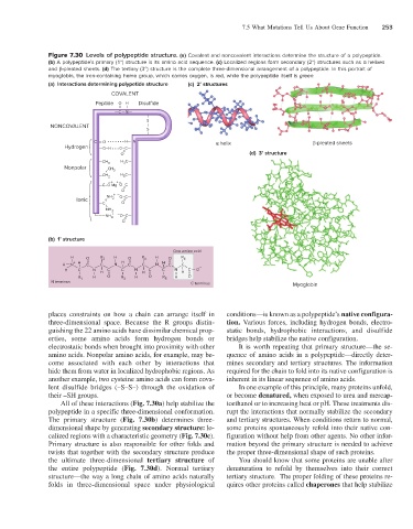
7.5 What Mutations Tell Us About Gene Function   253


                       Figure 7.30  Levels of polypeptide structure. (a) Covalent and noncovalent interactions determine the structure of a polypeptide.
                       (b) A polypeptide’s primary (1°) structure is its amino acid sequence. (c) Localized regions form secondary (2°) structures such as α helixes
                       and β-pleated sheets. (d) The tertiary (3°) structure is the complete three-dimensional arrangement of a polypeptide. In this portrait of
                       myoglobin, the iron-containing heme group, which carries oxygen, is red, while the polypeptide itself is green.
                       (a)  Interactions determining polypetide structure  (c)  2° structures
                                         COVALENT
                                     Peptide  = O  H I  Disulfide
                                          —C—N—
                                                    I
                                                    S
                       NONCOVALENT                  I
                                                    S
                                                    I
                                    C  = O  • • • • • • • H— N            helix                       -pleated sheets
                           Hydrogen   —O–H • • •O–C—
                                            O =                                   (d)  3° structure
                                      —CH 3  H C—
                                             3
                           Nonpolar     CH 3
                                       I
                                      —CH 2  H 3 C—
                                        –+  +–
                                      —C–O Mg  O–C—
                                             =
                                            O
                                        NH 2 +  – O–C—
                               Ionic    =    =
                                      —C    O
                                        I
                                        NH 2
                                    I ———NH +  – O–C—
                                         3    =
                                            O
                       (b)  1° structure
                                                            One amino acid
                             H    O   R 2  H  O   R 4  H  O   R 6
                             I  +  =  I   I  H  =  I   I   =  I
                           H     N  H I  C  C  N  I  C  C  N  H I  C  C
                             H          C  N  I  C  C  N  I  C  C  N  H I  C — O –
                                I   I  H  =  I  I  H  =  I  I    =
                               R 1  H   O   R 3  H  O   R 5  H  O
                       N terminus                                C terminus                    Myoglobin

                       places constraints on how a chain can arrange itself in     conditions—is known as a polypeptide’s native configura-
                       three-dimensional space. Because the R groups distin-  tion. Various forces, including hydrogen bonds, electro-
                       guishing the 22 amino acids have dissimilar chemical prop-  static bonds, hydrophobic interactions, and disulfide
                       erties, some amino acids form hydrogen bonds or     bridges help stabilize the native configuration.
                       electrostatic bonds when brought into proximity with other   It is worth repeating that primary structure—the se-
                       amino acids. Nonpolar amino acids, for example, may be-  quence of amino acids in a polypeptide—directly deter-
                       come associated with each other by interactions that   mines secondary and tertiary structures. The information
                       hide them from water in localized hydrophobic regions. As   required for the chain to fold into its native configuration is
                       another example, two cysteine amino acids can form cova-  inherent in its linear sequence of amino acids.
                       lent disulfide bridges (–S–S–) through the oxidation of   In one example of this principle, many proteins unfold,
                       their –SH groups.                                   or become denatured, when exposed to urea and mercap-
                          All of these interactions (Fig. 7.30a) help stabilize the   toethanol or to increasing heat or pH. These treatments dis-
                       polypeptide in a specific three-dimensional conformation.   rupt the interactions that normally stabilize the secondary
                       The primary structure (Fig. 7.30b) determines three-   and tertiary structures. When conditions return to normal,
                       dimensional shape by generating secondary structure: lo-  some proteins spontaneously refold into their native con-
                       calized regions with a characteristic geometry (Fig. 7.30c).   figuration without help from other agents. No other infor-
                       Primary structure is also responsible for other folds and   mation beyond the primary structure is needed to achieve
                       twists that together with the secondary structure produce   the proper three-dimensional shape of such proteins.
                       the  ultimate  three-dimensional  tertiary  structure  of    You should know that some proteins are unable after
                       the entire polypeptide (Fig. 7.30d). Normal tertiary    denaturation to refold by themselves into their correct
                       structure—the way a long chain of amino acids naturally   tertiary structure.  The proper folding of these proteins re-
                       folds in three-dimensional space under physiological   quires other proteins called chaperones that help stabilize