
Page 93 - Genetics_From_Genes_to_Genomes_6th_FULL_Part2
P. 93
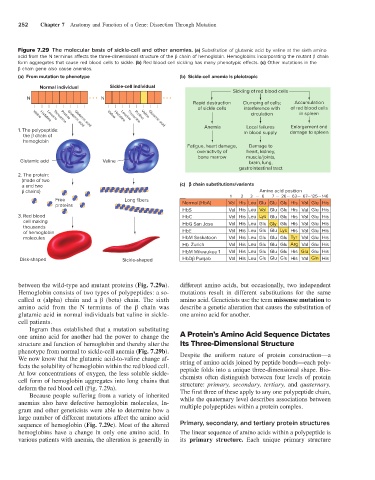
252    Chapter 7    Anatomy and Function of a Gene: Dissection Through Mutation


              Figure 7.29  The molecular basis of sickle-cell and other anemias. (a) Substitution of glutamic acid by valine at the sixth amino
              acid from the N terminus affects the three-dimensional structure of the β chain of hemoglobin. Hemoglobins incorporating the mutant β chain
              form aggregates that cause red blood cells to sickle. (b) Red blood cell sickling has many phenotypic effects. (c) Other mutations in the
              β chain gene also cause anemias.
              (a)  From mutation to phenotype                      (b)  Sickle-cell anemia is pleiotropic

                     Normal individual      Sickle-cell individual
                                                                                    Sickling of red blood cells
                 N                   ••• N                   •••
                                                                       Rapid destruction  Clumping of cells;  Accumulation
                                                                         of sickle cells  interference with  of red blood cells
                                                                                          circulation     in spleen
                                           Valine
                                                      Valine
                   Valine
                                                    Proline
                            Proline
                        Leucine
                                                Leucine
                     Histidine
                                              Histidine
                          Threonine
                                                  Threonine
                                                                           Anemia        Local failures  Enlargement and
                                 Glutamic acid
                              Glutamic acid
                                                         Glutamic acid
                                                         Glutamic acid
              1. The polypeptide:                                                       in blood supply  damage to spleen
                  the   chain of
                  hemoglobin
                                                                     Fatigue, heart damage,  Damage to
                                                                         overactivity of  heart, kidney,
                                                                         bone marrow     muscle/joints,
               Glutamic acid              Valine                                         brain, lung,
                                                                                      gastrointestinal tract
              2. The protein:
                 (made of two
                   and two                                         (c)      chain substitutions/variants
                   chains)                                                                   Amino acid position
                                                                                                             …
                                                                                   1   2  3 …  6  7 … 26 … 63  … 67 125  … 146
                          Free                   Long fibers
                          proteins                                  Normal (HbA)   Val His Leu Glu  Glu Glu  His  Val Glu  His
                                                                    HbS            Val His Leu  Val Glu  Glu  His  Val Glu  His
              3. Red blood                                          HbC            Val His Leu  Lys Glu  Glu  His  Val Glu  His
                  cell making                                       HbG San Jose   Val His Leu Glu Gly  Glu  His  Val Glu  His
                  thousands
                  of hemoglobin                                     HbE            Val His Leu Glu  Glu  Lys His  Val Glu  His
                  molecules                                         HbM Saskatoon  Val His Leu Glu  Glu  Glu Tyr  Val Glu  His
                                                                    Hb Zurich      Val His Leu Glu  Glu  Glu Arg  Val Glu  His
                                                                    HbM Milwaukee 1  Val His Leu Glu  Glu  Glu  His Glu Glu  His
               Disk-shaped                      Sickle-shaped       HbD  Punjab    Val His Leu Glu  Glu  Glu  His  Val Gln  His
              between the wild-type and mutant proteins (Fig. 7.29a).   different amino acids, but occasionally, two independent
              Hemoglobin consists of two types of polypeptides: a so-  mutations result in different substitutions for the same
              called α (alpha) chain and a β (beta) chain. The sixth   amino acid. Geneticists use the term missense mutation to
              amino  acid  from the  N  terminus  of  the  β  chain  was    describe a genetic alteration that causes the substitution of
              glutamic acid in normal individuals but valine in sickle-  one amino acid for another.
              cell patients.
                  Ingram thus established that a mutation substituting
              one amino acid for another had the power to change the   A Protein’s Amino Acid Sequence Dictates
              structure and function of hemoglobin and thereby alter the   Its Three-Dimensional Structure
              phenotype from normal to sickle-cell anemia (Fig. 7.29b).   Despite the uniform nature of protein construction—a
              We now know that the glutamic acid-to-valine change af-  string of amino acids joined by peptide bonds—each poly-
              fects the solubility of hemoglobin within the red blood cell.   peptide folds into a unique three-dimensional shape. Bio-
              At low concentrations of oxygen, the less soluble sickle-  chemists often distinguish between four levels of protein
              cell form of hemoglobin aggregates into long chains that   structure:  primary, secondary, tertiary, and  quaternary.
              deform the red blood cell (Fig. 7.29a).              The first three of these apply to any one polypeptide chain,
                  Because people suffering from a variety of inherited
              anemias also have defective hemoglobin molecules, In-  while the quaternary level describes associations between
                                                                   multiple polypeptides within a protein complex.
              gram and other geneticists were able to determine how a
              large number of different mutations affect the amino acid
              sequence of hemoglobin (Fig. 7.29c). Most of the altered   Primary, secondary, and tertiary protein structures
              hemoglobins have a change in only one amino acid. In   The linear sequence of amino acids within a polypeptide is
                various patients with anemia, the alteration is generally in   its  primary structure. Each unique primary structure