
Page 134 - Genetics_From_Genes_to_Genomes_6th_FULL_Part2
P. 134
8.3 Translation: From mRNA to Protein 293
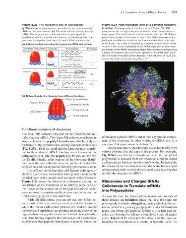
Figure 8.23 The ribosome: Site of polypeptide Figure 8.24 High resolution view of a bacterial ribosome
synthesis. (a) A ribosome has two subunits, each composed of in action. The large subunit is at the top; its 23S and 5S rRNA
rRNA and various proteins. (b) The small subunit initially binds to components are in bright blue and its various protein components in
mRNA. The large subunit contributes the enzyme peptidyl bright green. The small subunit is at the bottom, with the 16S rRNA in
transferase, which catalyzes the formation of peptide bonds. The gray and its protein components in aqua. Two tRNA molecules are in
two subunits together form the A, P, and E tRNA binding sites. gold, with the tRNA on the left at the E site and the tRNA to its right
(a) A ribosome has two subunits composed of RNA and protein. at the P site. The A site is occupied by a protein release factor shown
in pink. A few of the nucleotides in the mRNA (red) can be seen near
Complete Ribosomes Subunits Nucleotides Proteins the bottoms of the tRNAs and release factor. This ribosome is acting during
Prokaryotic a stage of translation just prior to the ejection of the tRNA from the E
31 site during the termination phase depicted in the left panel of Fig. 8.25c.
50S 23S rRNA 3000 nucleotides
© Yuxin Mao, Ph.D., Cornell University, Ithaca, NY
5S rRNA 120 nucleotides
70S 21
30S 16S rRNA 1700 nucleotides
28S rRNA
Eukaryotic
5000 nucleotides
~ 45
5.8S rRNA 5S rRNA
160 nucleotides 120 nucleotides
60S
80S 40S ~ 33
18S rRNA 2000 nucleotides
(b) Di erent parts of a ribosome have di erent functions.
Peptidyl transferase
Peptidyl (P) site
Aminoacyl (A) site
Large subunit
Exit (E) site
Small subunit
Functional domains of ribosomes
The small 30S subunit is the part of the ribosome that ini
tially binds to mRNA. The larger 50S subunit contributes an of the large subunit’s rRNA rather than any protein compo
enzyme known as peptidyl transferase, which catalyzes nent of the ribosome. In other words, the rRNA acts as a
formation of the peptide bonds joining adjacent amino acids ribozyme that joins amino acids together.
(Fig. 8.23b). Both the small and the large subunits contrib During translation, the ribosome associates briefly with
ute to three distinct tRNA binding areas known as the various proteins that aid steps in the process. For example,
aminoacyl (or A) site, the peptidyl (or P) site, and the exit Fig. 8.24 shows that late in translation when the completed
(or E) site. Finally, other regions of the ribosome distrib polypeptide is released from the ribosome, a protein called
uted over the two subunits serve as points of contact for a release factor binds to the ribosome’s A site. Remarkably,
some of the additional proteins that play roles in translation. the release factor can associate with the A site because part
Using Xray crystallography and elegant techniques of of this protein folds in threedimensional space in a way that
electron microscopy, researchers have gained a remarkably mimics the structure of a tRNA.
detailed view of the complicated structure of the ribosome.
Figure 8.24 shows the interior of a ribosome nearing the Ribosomes and Charged tRNAs
completion of the translation of an mRNA; some parts of Collaborate to Translate mRNAs
the ribosome that extend out of the page toward the reader into Polypeptides
were removed computationally so you can better see the
tRNAs occupying the E site and P site. As was the case for transcription, translation consists of
With this illustration, you can see that the rRNAs oc three phases: an initiation phase that sets the stage for
cupy most of the space in the central part of the ribosome, polypeptide synthesis; elongation, during which amino ac
while the various ribosomal proteins are studded around ids are added to a growing polypeptide; and a termination
the exterior. Surprisingly, no proteins are found close to the phase that brings polypeptide synthesis to a halt and en
region where the peptide bonds are formed during transla ables the ribosome to release a completed chain of amino
tion. This finding supports the conclusions of biochemical acids. Figure 8.25 illustrates the details of the process,
experiments that peptidyl transferase is actually a function focusing on translation as it occurs in bacterial cells. As