
Page 99 - Genetics_From_Genes_to_Genomes_6th_FULL_Part1
P. 99
4.1 Chromosomes: The Carriers of Genes 91
Genes Reside in Chromosomes fertilization forms the zygote, the process of mitosis then
ensures that all the somatic cells of the developing individ-
Further investigations, some dependent on technical inno- ual have identical diploid chromosome sets.
vations in microscopy, suggested that yet smaller, discrete
structures within the nucleus are the repository of genetic Species variations in the number
information. In the 1880s, for example, a newly discovered and shape of chromosomes
combination of organic and inorganic dyes revealed the
existence of the long, brightly staining, threadlike bodies Scientists analyze the chromosomal makeup of a cell when
within the nucleus that we call chromosomes (literally col- the chromosomes are most visible—at a specific moment
ored bodies). It was now possible to follow the movement in the cell cycle of growth and division, just before the
of chromosomes during different kinds of cell division. nucleus divides. At this point, known as metaphase
In embryonic cells, the chromosomal threads split (described in detail later), individual chromosomes have
lengthwise in two just before cell division, and each of the duplicated and condensed from thin threads into compact
two newly forming daughter cells receives one-half of rodlike structures. Each chromosome now consists of two
every split thread. The kind of nuclear division followed by identical halves known as sister chromatids (Fig. 4.3).
cell division that results in two daughter cells containing The specific location at which sister chromatids are
the same number and type of chromosomes as the original attached to each other is called the centromere. Each sister
parent cell is called mitosis (from the Greek mitos meaning chromatid has its own centromere (Fig. 4.3), but in the du-
thread and -osis meaning formation or increase). plicated chromosome, the two sister centromeres are pulled
In the cells that give rise to male and female gametes, together so tightly that they form a constriction within
the chromosomes composing each pair become segregated, which they cannot be resolved from each other, even in
so that the resulting gametes receive only one chromosome images obtained in the scanning electron microscope (see
from each chromosome pair. The kind of nuclear division the picture at the beginning of the chapter).
that generates egg or sperm cells containing half the number
of chromosomes found in other cells within the same organ-
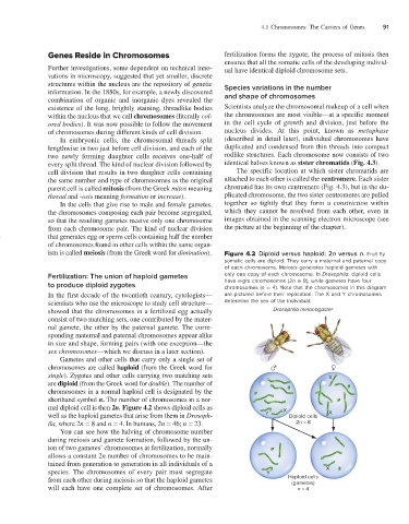
ism is called meiosis (from the Greek word for diminution). Figure 4.2 Diploid versus haploid: 2n versus n. Fruit fly
somatic cells are diploid: They carry a maternal and paternal copy
of each chromosome. Meiosis generates haploid gametes with
Fertilization: The union of haploid gametes only one copy of each chromosome. In Drosophila, diploid cells
to produce diploid zygotes have eight chromosomes (2n = 8), while gametes have four
chromosomes (n = 4). Note that the chromosomes in this diagram
In the first decade of the twentieth century, cytologists— are pictured before their replication. The X and Y chromosomes
scientists who use the microscope to study cell structure— determine the sex of the individual.
showed that the chromosomes in a fertilized egg actually Drosophila melanogaster
consist of two matching sets, one contributed by the mater-
nal gamete, the other by the paternal gamete. The corre-
sponding maternal and paternal chromosomes appear alike
in size and shape, forming pairs (with one exception—the
sex chromosomes—which we discuss in a later section).
Gametes and other cells that carry only a single set of
chromosomes are called haploid (from the Greek word for
single). Zygotes and other cells carrying two matching sets X
are diploid (from the Greek word for double). The number of
chromosomes in a normal haploid cell is designated by the
shorthand symbol n. The number of chromosomes in a nor-
mal diploid cell is then 2n. Figure 4.2 shows diploid cells as X
well as the haploid gametes that arise from them in Drosoph- Y Diploid cells X
ila, where 2n = 8 and n = 4. In humans, 2n = 46; n = 23. 2n = 8
You can see how the halving of chromosome number
during meiosis and gamete formation, followed by the un-
ion of two gametes’ chromosomes at fertilization, normally
allows a constant 2n number of chromosomes to be main-
tained from generation to generation in all individuals of a
species. The chromosomes of every pair must segregate Y X
from each other during meiosis so that the haploid gametes Haploid cells
(gametes)
will each have one complete set of chromosomes. After n = 4