
Page 100 - Genetics_From_Genes_to_Genomes_6th_FULL_Part1
P. 100
92 Chapter 4 The Chromosome Theory of Inheritance
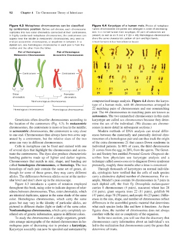
Figure 4.3 Metaphase chromosomes can be classified Figure 4.4 Karyotype of a human male. Photos of metaphase
by centromere position. Before cell division, each chromosome human chromosomes are paired and arranged in order of decreasing
replicates into two sister chromatids connected at their centromeres. size. In a normal human male karyotype, 22 pairs of autosomes are
In highly condensed metaphase chromosomes, the centromeres can present, as well as an X and a Y (2n = 46). Homologous chromosomes
appear near the middle (a metacentric chromosome), very near an share the same characteristic pattern of dark and light bands.
end (an acrocentric chromosome), or anywhere in between. In a © Scott Camazine & Sue Trainor/Science Source
diploid cell, one homologous chromosome in each pair is from the
mother and the other from the father.
Pair of Homologous Pair of Homologous
Metacentric Chromosomes Acrocentric Chromosomes
Centromere Centromere
Sister Nonsister
chromatids chromatids
Nonhomologous chromosomes computerized image analysis. Figure 4.4 shows the karyo-
type of a human male, with 46 chromosomes arranged in
22 matching pairs of chromosomes and one nonmatching
Homologous chromosomes Homologous chromosomes
pair. The 44 chromosomes in matching pairs are known as
autosomes. The two unmatched chromosomes in this male
Geneticists often describe chromosomes according to karyotype are called sex chromosomes because they deter-
the location of the centromere (Fig. 4.3). In metacentric mine the sex of the individual. (We discuss sex chromo-
chromosomes, the centromere is more or less in the middle; somes in more detail in subsequent sections.)
in acrocentric chromosomes, the centromere is very close Modern methods of DNA analysis can reveal differ-
to one end. Chromosomes thus always have two arms sep- ences between the maternally and paternally derived chro-
arated by a centromere, but the relative sizes of the two mosomes of a homologous pair and can thus track the origin
arms can vary in different chromosomes. of the extra chromosome 21 that causes Down syndrome in
Cells in metaphase can be fixed and stained with one individual patients. In 80% of cases, the third chromosome
of several dyes that highlight the chromosomes and accen- 21 comes from the egg; in 20%, from the sperm. The Genet-
tuate the centromeres. The dyes also produce characteristic ics and Society box entitled Prenatal Genetic Diagnosis de-
banding patterns made up of lighter and darker regions. scribes how physicians use karyotype analysis and a
Chromosomes that match in size, shape, and banding are technique called amniocentesis to diagnose Down syndrome
called homologous chromosomes, or homologs. The two prenatally, roughly three months after a fetus is conceived.
homologs of each pair contain the same set of genes, al- Through thousands of karyotypes on normal individu-
though for some of those genes, they may carry different als, cytologists have verified that the cells of each species
alleles. The differences between alleles occur at the molec- carry a distinctive diploid number of chromosomes. For ex-
ular level and don’t show up in the microscope. ample, Mendel’s peas contain 14 chromosomes in 7 pairs in
Figure 4.3 introduces a system of notation employed each diploid cell, the fruit fly Drosophila melanogaster
throughout this book, using color to indicate degrees of relat- carries 8 chromosomes (4 pairs), macaroni wheat has 28
edness between chromosomes. Thus, sister chromatids, which (14 pairs), giant sequoia trees 22 (11 pairs), goldfish 94
are identical duplicates, appear in the same shade of the same (47 pairs), dogs 78 (39 pairs), and people 46 (23 pairs). Differ-
color. Homologous chromosomes, which carry the same ences in the size, shape, and number of chromosomes reflect
genes but may vary in the identity of particular alleles, are differences in the assembled genetic material that determines
pictured in different shades (light or dark) of the same color. what each species looks like and how it functions. As these
Nonhomologous chromosomes, which carry completely un- figures show, the number of chromosomes does not always
related sets of genetic information, appear in different colors. correlate with the size or complexity of the organism.
To study the chromosomes of a single organism, geneti- In the next section, you will see that the discovery that
cists arrange micrographs of the stained chromosomes in ho- chromosomes carry information about an individual’s sex
mologous pairs of decreasing size to produce a karyotype. led to the realization that chromosomes carry the genes that
Karyotype assembly can now be speeded and automated by determine all traits.