
Page 101 - Genetics_From_Genes_to_Genomes_6th_FULL_Part1
P. 101
4.1 Chromosomes: The Carriers of Genes 93
GENETICS AND SOCIETY Crowd: © Image Source/Getty Images RF
Prenatal Genetic Diagnosis
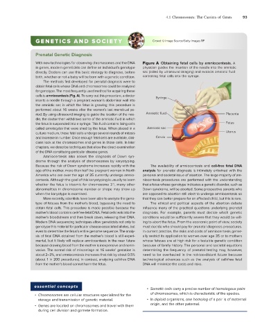
With new technologies for observing chromosomes and the DNA Figure A Obtaining fetal cells by amniocentesis. A
in genes, modern geneticists can define an individual’s genotype physician guides the insertion of the needle into the amniotic
directly. Doctors can use this basic strategy to diagnose, before sac (aided by ultrasound imaging) and extracts amniotic fluid
birth, whether or not a baby will be born with a genetic condition. containing fetal cells into the syringe.
The methods first developed for prenatal diagnosis were to
obtain fetal cells whose DNA and chromosomes could be analyzed
for genotype. The most frequently used method for acquiring these
cells is amniocentesis (Fig. A). To carry out this procedure, a doctor Syringe
inserts a needle through a pregnant woman’s abdominal wall into
the amniotic sac in which the fetus is growing; this procedure is
performed about 16 weeks after the woman’s last menstrual pe-
riod. By using ultrasound imaging to guide the location of the nee- Amniotic fluid Placenta
dle, the doctor then withdraws some of the amniotic fluid in which
the fetus is suspended into a syringe. This fluid contains living cells Fetus
called amniocytes that were shed by the fetus. When placed in a Amniotic sac
culture medium, these fetal cells undergo several rounds of mitosis Uterus
and increase in number. Once enough fetal cells are available, clini- Cervix
cians look at the chromosomes and genes in those cells. In later
chapters, we describe techniques that allow the direct examination
of the DNA constituting particular disease genes.
Amniocentesis also allows the diagnosis of Down syn-
drome through the analysis of chromosomes by karyotyping.
Because the risk of Down syndrome increases rapidly with the The availability of amniocentesis and cell-free fetal DNA
age of the mother, more than half the pregnant women in North analysis for prenatal diagnosis is intimately entwined with the
America who are over the age of 35 currently undergo amnio- personal and societal issue of abortion. The large majority of am-
centesis. Although the goal of this karyotyping is usually to learn niocentesis procedures are performed with the understanding
whether the fetus is trisomic for chromosome 21, many other that a fetus whose genotype indicates a genetic disorder, such as
abnormalities in chromosome number or shape may show up Down syndrome, will be aborted. Some prospective parents who
when the karyotype is examined. are opposed to abortion still elect to undergo amniocentesis so
More recently, scientists have been able to analyze the geno- that they can better prepare for an affected child, but this is rare.
type of fetuses from the mother’s blood, bypassing the need to The ethical and political aspects of the abortion debate
obtain fetal cells. This procedure is made possible because the influence many of the practical questions underlying prenatal
mother’s blood contains cell-free fetal DNA. Fetal cells leak into the diagnosis. For example, parents must decide which genetic
mother’s bloodstream and then break down, releasing their DNA. conditions would be sufficiently severe that they would be will-
Modern DNA sequencing techniques allow geneticists not only to ing to abort the fetus. From the economic point of view, society
genotype this material for particular disease-associated alleles, but must decide who should pay for prenatal diagnosis procedures.
even to determine the fetus’s entire genome sequence. The analy- In current practice, the risks and costs of amniocentesis gener-
sis of fetal DNA obtained from the mother’s blood is still experi- ally restrict its application to women over age 35 or to mothers
mental, but it likely will replace amniocentesis in the near future whose fetuses are at high risk for a testable genetic condition
because drawing blood from the mother is inexpensive and nonin- because of family history. The personal and societal equations
vasive. The normal risk of miscarriage at 16 weeks’ gestation is determining the frequency of prenatal testing may, however,
about 2–3%, and amniocentesis increases that risk by about 0.5% need to be overhauled in the not-too-distant future because
(about 1 in 200 precedures). In contrast, analyzing cell-free DNA technological advances such as the analysis of cell-free fetal
from the mother’s blood cannot harm the fetus. DNA will minimize the costs and risks.
essential concepts
• Somatic cells carry a precise number of homologous pairs
• Chromosomes are cellular structures specialized for the of chromosomes, which is characteristic of the species.
storage and transmission of genetic material. • In diploid organisms, one homolog of a pair is of maternal
• Genes are located on chromosomes and travel with them origin, and the other paternal.
during cell division and gamete formation.