
Page 106 - Genetics_From_Genes_to_Genomes_6th_FULL_Part1
P. 106
98 Chapter 4 The Chromosome Theory of Inheritance
4.3 Mitosis: Cell Division That eventually ends up enclosed in a separate nucleus in a sepa-
rate cell. The two cells, known as daughter cells, are thus
Preserves Chromosome Number genetically identical.
The repeating pattern of cell growth (an increase in
size) followed by division (the splitting of one cell into
learning objectives two) is called the cell cycle (Fig. 4.9). Only a small part of
the cell cycle is spent in division (or M phase); the period
1. Describe the key chromosome behaviors during mitosis. between divisions is called interphase.
2. Diagram the forces and structures that dictate
chromosomal movement during mitosis.
During Interphase, Cells Grow and
Replicate Their Chromosomes
The fertilized human egg is a single diploid cell that pre- Interphase consists of three parts: gap 1 (G 1 ), synthesis (S),
serves its genetic identity unchanged through more than and gap 2 (G 2 ) (Fig. 4.9). G 1 lasts from the birth of a new cell
100 generations of cells as it divides again and again to to the onset of chromosome replication; for the genetic mate-
produce a full-term infant ready to be born. As the newborn rial, it is a period when the chromosomes are neither dupli-
infant develops into a toddler, a teenager, and an adult, yet cating nor dividing. During this time, the cell achieves most
more cell divisions fuel continued growth and maturation. of its growth by using the information from its genes to make
Mitosis, the nuclear division that apportions chromosomes and assemble the materials it needs to function normally. G 1
in equal fashion to two daughter cells, is the cellular mech- varies in length more than any other phase of the cell cycle.
anism that preserves genetic information through all these In rapidly dividing cells of the human embryo, for example,
generations of cells. In this section, we take a close look at G 1 is as short as a few hours. In contrast, mature brain cells
how the nuclear division of mitosis fits into the overall
scheme of cell growth and division.
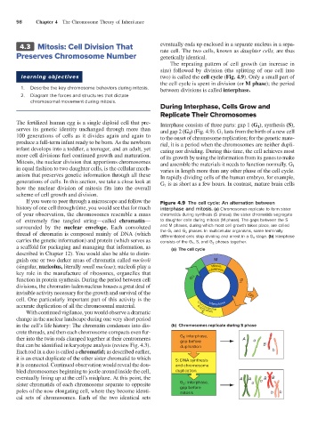
If you were to peer through a microscope and follow the Figure 4.9 The cell cycle: An alternation between
history of one cell through time, you would see that for much interphase and mitosis. (a) Chromosomes replicate to form sister
of your observation, the chromosomes resemble a mass chromatids during synthesis (S phase); the sister chromatids segregate
of extremely fine tangled string—called chromatin— to daughter cells during mitosis (M phase). The gaps between the S
surrounded by the nuclear envelope. Each convoluted and M phases, during which most cell growth takes place, are called
thread of chromatin is composed mainly of DNA (which the G 1 and G 2 phases. In multicellular organisms, some terminally
differentiated cells stop dividing and arrest in a G 0 stage. (b) Interphase
carries the genetic information) and protein (which serves as consists of the G 1 , S, and G 2 phases together.
a scaffold for packaging and managing that information, as (a) The cell cycle
described in Chapter 12). You would also be able to distin-
guish one or two darker areas of chromatin called nucleoli M
(singular, nucleolus, literally small nucleus); nucleoli play a Mitosis,
key role in the manufacture of ribosomes, organelles that cytokinesis
function in protein synthesis. During the period between cell G 2 G 1
divisions, the chromatin-laden nucleus houses a great deal of
invisible activity necessary for the growth and survival of the G
cell. One particularly important part of this activity is the Interphase 0
accurate duplication of all the chromosomal material. duplication
Chromosome
With continued vigilance, you would observe a dramatic S
change in the nuclear landscape during one very short period
in the cell’s life history: The chromatin condenses into dis- (b) Chromosomes replicate during S phase
crete threads, and then each chromosome compacts even fur-
ther into the twin rods clamped together at their centromeres G : interphase, A a
1
gap before
that can be identified in karyotype analysis (review Fig. 4.3). duplication B
Each rod in a duo is called a chromatid; as described earlier, b
it is an exact duplicate of the other sister chromatid to which S: DNA synthesis
it is connected. Continued observation would reveal the dou- and chromosome
bled chromosomes beginning to jostle around inside the cell, duplication
eventually lining up at the cell’s midplane. At this point, the A A a B B
sister chromatids of each chromosome separate to opposite G : interphase, a
2
poles of the now elongating cell, where they become identi- gap before b b
mitosis
cal sets of chromosomes. Each of the two identical sets