
Page 31 - Genetics_From_Genes_to_Genomes_6th_FULL_Part2
P. 31
190 Chapter 6 DNA Structure, Replication, and Recombination
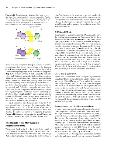
Figure 6.10 Complementary base pairing. An A on one exist,” the beauty of the structure is not necessarily evi-
strand can form two (noncovalent) hydrogen bonds with a T on the dence of its correctness. At the time of its presentation, the
other strand. G on one strand can form three hydrogen bonds with strongest evidence for its correctness was its physical plau-
a C on the other strand. The size and shape of A–T and of G–C sibility, its chemical and spatial compatibility with all
base pairs are similar, allowing both to fill the same amount of
space between the two backbones of the double helix. available data, and its capacity for explaining many bio-
Hydrogen bonds logical phenomena.
G H C
N O H N B DNA and Z DNA
The majority of naturally occurring DNA molecules have
N N H N the configuration suggested by Watson and Crick. Such
Sugar N N molecules are known as B-form DNA; they spiral to the
N H O
Sugar right (Fig. 6.12a). DNA structure is, however, more poly-
morphic than originally assumed. One type, for example,
A H T contains nucleotide sequences that cause the DNA to as-
sume what is known as a Z form in which the helix spi-
N N H O CH 3
rals to the left and the backbone takes on a zigzag shape
N N H N (Fig. 6.12b). Researchers have observed many kinds of
Sugar N N unusual non-B structures in vitro (in the test tube, literally
Hydrogen bonds O Sugar in glass), and they speculate that some of these might oc-
cur at least transiently in living cells. There is some evi-
dence, for instance, that Z DNA might exist in certain
atoms on purines and pyrimidines play a crucial role in mo- chromosomal regions in vivo (in the living organism).
lecular interactions as they can participate in the formation Whether the Z form and other unusual conformations
of hydrogen bonds: weak electrostatic bonds that result in have any biological role remains to be determined.
a partial sharing of hydrogen atoms between reacting groups
(Fig. 6.10). Watson saw that A and T could be paired to- Linear and circular DNA
gether such that two hydrogen bonds formed between them. The nuclear chromosomes of all eukaryotic organisms are
If G and C were similarly paired, hydrogen bonds could also long, linear double helixes, but some smaller chromosomes
easily connect the nucleotides carrying these two bases. are circular (Fig. 6.13a and b). These include the chromo-
(Watson originally posited two hydrogen bonds between G somes of prokaryotic bacteria, the chromosomes of organ-
and C, but there are actually three.) Remarkably, the two elles such as the mitochondria and chloroplasts that are
pairs—A–T and G–C—had essentially the same shape. found inside eukaryotic cells, and the chromosomes of
This meant that the two pairs could fit in any order between some viruses, including the papovaviruses that can cause
two sugar-phosphate backbones without distorting the cancers in animals and humans. Such circular chromo-
structure. This complementary base pairing also ex- somes consist of covalently closed, double-stranded circu-
plained the Chargaff ratios—always equal amounts of A lar DNA molecules. Although neither strand of these
and T, and of G and C. Note that both of these base pairs circular double helixes has an end, the two strands are still
consist of one purine and one pyrimidine. antiparallel in polarity.
Crick connected the chemical facts with the X-ray
data, recognizing that because of the geometry of the base- Single-stranded and double-stranded DNA
sugar bonds in nucleotides, the orientation of the bases in
Watson’s pairing scheme could arise only if the bases were In some viruses, the genetic material consists of relatively
attached to backbones running in opposite directions. small, single-stranded DNA molecules. Once inside a cell,
Figure 6.11 illustrates and explains the model Watson and the single strand serves as a template (pattern) for making
Crick proposed in April 1953: DNA as a double helix. a second strand, and the resulting double-stranded DNA
then governs the production of more virus particles. Ex-
amples of viruses carrying single-stranded DNA are bacte-
The Double Helix May Assume riophages ϕX174 and M13, and mammalian parvoviruses,
Alternative Forms which are associated with fetal death and spontaneous
abortion in humans. In both ϕX174 and M13, the single
Watson and Crick arrived at the double helix model of DNA strand is in the form of a covalently closed circle; in
DNA structure by building models, not by a direct struc- the parvoviruses, it is linear (Fig. 6.13c and d).
tural determination from the data alone. And even though Alternative B and Z configurations; circularization of the
Watson has written that “a structure this pretty just had to molecule; and single strands that are converted to double