
Page 24 - Genetics_From_Genes_to_Genomes_6th_FULL_Part2
P. 24
6.1 Experimental Evidence for DNA as the Genetic Material 183
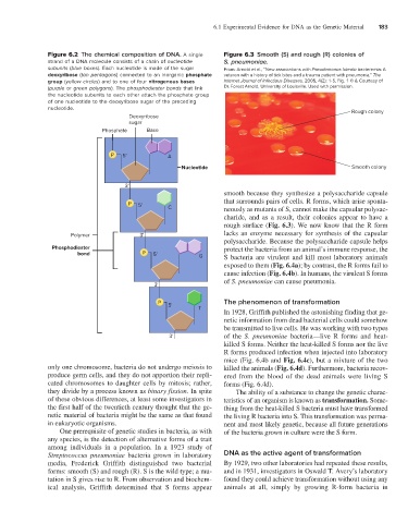
Figure 6.2 The chemical composition of DNA. A single Figure 6.3 Smooth (S) and rough (R) colonies of
strand of a DNA molecule consists of a chain of nucleotide S. pneumoniae.
subunits (blue boxes). Each nucleotide is made of the sugar From: Arnold et al., “New associations with Pseudomonas luteola bacteremia: A
deoxyribose (tan pentagons) connected to an inorganic phosphate veteran with a history of tick bites and a trauma patient with pneumonia,” The
group (yellow circles) and to one of four nitrogenous bases Internet Journal of Infectious Diseases, 2005, 4(2): 1-5, Fig. 1 © & Courtesy of
(purple or green polygons). The phosphodiester bonds that link Dr. Forest Arnold, University of Louisville. Used with permission.
the nucleotide subunits to each other attach the phosphate group
of one nucleotide to the deoxyribose sugar of the preceding
nucleotide. Rough colony
Deoxyribose
sugar
Phosphate Base
P 5' A
Nucleotide Smooth colony
3'
smooth because they synthesize a polysaccharide capsule
that surrounds pairs of cells. R forms, which arise sponta-
P 5'
C neously as mutants of S, cannot make the capsular polysac-
charide, and as a result, their colonies appear to have a
rough surface (Fig. 6.3). We now know that the R form
Polymer 3' lacks an enzyme necessary for synthesis of the capsular
polysaccharide. Because the polysaccharide capsule helps
Phosphodiester protect the bacteria from an animal’s immune response, the
bond P 5' G S bacteria are virulent and kill most laboratory animals
exposed to them (Fig. 6.4a); by contrast, the R forms fail to
cause infection (Fig. 6.4b). In humans, the virulent S forms
of S. pneumoniae can cause pneumonia.
3'
P The phenomenon of transformation
5'
T
In 1928, Griffith published the astonishing finding that ge-
netic information from dead bacterial cells could somehow
be transmitted to live cells. He was working with two types
3' of the S. pneumoniae bacteria—live R forms and heat-
killed S forms. Neither the heat-killed S forms nor the live
R forms produced infection when injected into laboratory
mice (Fig. 6.4b and Fig. 6.4c), but a mixture of the two
only one chromosome, bacteria do not undergo meiosis to killed the animals (Fig. 6.4d). Furthermore, bacteria recov-
produce germ cells, and they do not apportion their repli- ered from the blood of the dead animals were living S
cated chromosomes to daughter cells by mitosis; rather, forms (Fig. 6.4d).
they divide by a process known as binary fission. In spite The ability of a substance to change the genetic charac-
of these obvious differences, at least some investigators in teristics of an organism is known as transformation. Some-
the first half of the twentieth century thought that the ge- thing from the heat-killed S bacteria must have transformed
netic material of bacteria might be the same as that found the living R bacteria into S. This transformation was perma-
in eukaryotic organisms. nent and most likely genetic, because all future generations
One prerequisite of genetic studies in bacteria, as with of the bacteria grown in culture were the S form.
any species, is the detection of alternative forms of a trait
among individuals in a population. In a 1923 study of
Streptococcus pneumoniae bacteria grown in laboratory DNA as the active agent of transformation
media, Frederick Griffith distinguished two bacterial By 1929, two other laboratories had repeated these results,
forms: smooth (S) and rough (R). S is the wild type; a mu- and in 1931, investigators in Oswald T. Avery’s laboratory
tation in S gives rise to R. From observation and biochem- found they could achieve transformation without using any
ical analysis, Griffith determined that S forms appear animals at all, simply by growing R-form bacteria in