
Page 26 - Genetics_From_Genes_to_Genomes_6th_FULL_Part3
P. 26
320 Chapter 9 Digital Analysis of DNA
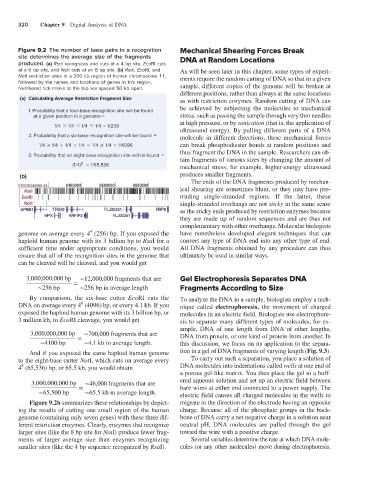
Figure 9.2 The number of base pairs in a recognition Mechanical Shearing Forces Break
site determines the average size of the fragments DNA at Random Locations
produced. (a) RsaI recognizes and cuts at a 4 bp site, EcoRI cuts
at a 6 bp site, and NotI cuts at an 8 bp site. (b) RsaI, EcoRI, and As will be seen later in this chapter, some types of experi-
NotI restriction sites in a 200 kb region of human chromosome 11, ments require the random cutting of DNA so that in a given
followed by the names and locations of genes in this region.
Numbered tick marks at the top are spaced 50 kb apart. sample, different copies of the genome will be broken at
different positions, rather than always at the same locations
(a) Calculating Average Restriction Fragment Size as with restriction enzymes. Random cutting of DNA can
be achieved by subjecting the molecules to mechanical
1. Probability that a four-base recognition site will be found
at a given position in a genome = stress, such as passing the sample through very thin needles
at high pressure, or by sonication (that is, the application of
1/4 1/4 1/4 1/4 = 1/256
ultrasound energy). By pulling different parts of a DNA
2. Probability that a six-base recognition site will be found = molecule in different directions, these mechanical forces
1/4 1/4 1/4 1/4 1/4 1/4 = 1/4096 can break phosphodiester bonds at random positions and
thus fragment the DNA in the sample. Researchers can ob-
3. Probability that an eight-base recognition site will be found =
tain fragments of various sizes by changing the amount of
8
(1/4) = 1/65,536 mechanical stress; for example, higher-energy ultrasound
(b) produces smaller fragments.
The ends of the DNA fragments produced by mechan-
ical shearing are sometimes blunt, or they may have pro-
truding single-stranded regions. If the latter, these
single-stranded overhangs are not sticky in the same sense
as the sticky ends produced by restriction enzymes because
they are made up of random sequences and are thus not
complementary with other overhangs. Molecular biologists
4
genome on average every 4 (256) bp. If you exposed the have nonetheless developed elegant techniques that can
haploid human genome with its 3 billion bp to RsaI for a convert any type of DNA end into any other type of end.
sufficient time under appropriate conditions, you would All DNA fragments obtained by any procedure can thus
ensure that all of the recognition sites in the genome that ultimately be used in similar ways.
can be cleaved will be cleaved, and you would get
3,000,000,000 bp ∼12,000,000 fragments that are Gel Electrophoresis Separates DNA
∼256 bp = ∼256 bp in average length Fragments According to Size
By comparison, the six-base cutter EcoRI cuts the To analyze the DNA in a sample, biologists employ a tech-
6
DNA on average every 4 (4096) bp, or every 4.1 kb. If you nique called electrophoresis, the movement of charged
exposed the haploid human genome with its 3 billion bp, or molecules in an electric field. Biologists use electrophore-
3 million kb, to EcoRI cleavage, you would get sis to separate many different types of molecules, for ex-
ample, DNA of one length from DNA of other lengths,
3,000,000,000 bp ∼700,000 fragments that are DNA from protein, or one kind of protein from another. In
∼4100 bp = ∼4.1 kb in average length. this discussion, we focus on its application to the separa-
And if you exposed the same haploid human genome tion in a gel of DNA fragments of varying length (Fig. 9.3).
to the eight-base cutter NotI, which cuts on average every To carry out such a separation, you place a solution of
8
4 (65,536) bp, or 65.5 kb, you would obtain DNA molecules into indentations called wells at one end of
a porous gel-like matrix. You then place the gel in a buff-
3,000,000,000 bp ∼46,000 fragments that are ered aqueous solution and set up an electric field between
∼65,500 bp = ∼65.5 kb in average length. bare wires at either end connected to a power supply. The
electric field causes all charged molecules in the wells to
Figure 9.2b summarizes these relationships by depict- migrate in the direction of the electrode having an opposite
ing the results of cutting one small region of the human charge. Because all of the phosphate groups in the back-
genome (containing only seven genes) with these three dif- bone of DNA carry a net negative charge in a solution near
ferent restriction enzymes. Clearly, enzymes that recognize neutral pH, DNA molecules are pulled through the gel
larger sites (like the 8 bp site for NotI) produce fewer frag- toward the wire with a positive charge.
ments of larger average size than enzymes recognizing Several variables determine the rate at which DNA mole-
smaller sites (like the 4 bp sequence recognized by RsaI). cules (or any other molecules) move during electrophoresis.