
Page 45 - Genetics_From_Genes_to_Genomes_6th_FULL_Part2
P. 45
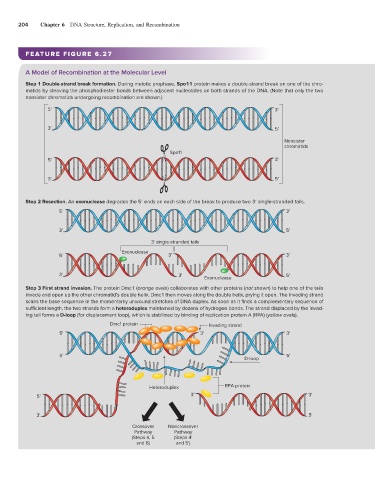
204    Chapter 6    DNA Structure, Replication, and Recombination


                 FEATURE FIGURE 6.27


                 A Model of Recombination at the Molecular Level
                 Step 1 Double-strand break formation. During meiotic prophase, Spo11 protein makes a double-strand break on one of the chro-
                 matids by cleaving the phosphodiester bonds between adjacent nucleotides on both strands of the DNA. (Note that only the two
                 nonsister chromatids undergoing recombination are shown.)
                        5'                                                                        3'


                        3'                                                                        5'

                                                                                                     Nonsister
                                                                                                     chromatids
                                                                Spo11
                        5'                                                                        3'

                        3'                                                                        5'


                 Step 2 Resection. An exonuclease degrades the 5′ ends on each side of the break to produce two 3′ single-stranded tails.
                            5'                                                                        3'


                            3'                                                                        5'
                                                          3' single-stranded tails
                                                Exonuclease
                            5'                                 3'                                     3'


                            3'                                     3'      Exonuclease                5'

                 Step 3 First strand invasion. The protein Dmc1 (orange ovals) collaborates with other proteins (not shown) to help one of the tails
                 invade and open up the other chromatid’s double helix. Dmc1 then moves along the double helix, prying it open. The invading strand
                 scans the base sequence in the momentarily unwound stretches of DNA duplex. As soon as it finds a complementary sequence of
                 sufficient length, the two strands form a heteroduplex maintained by dozens of hydrogen bonds. The strand displaced by the invad-
                 ing tail forms a D-loop (for displacement loop), which is stabilized by binding of replication protein A (RPA) (yellow ovals).
                                            Dmc1 protein                    Invading strand
                            5'                                            3'                          3'


                            3'                                                                        5'
                                                                                        D-loop


                                                         Heteroduplex             RPA protein
                    5'                                                3'                                     3'


                    3'                                                                                       5'
                                                   Crossover   Noncrossover
                                                    Pathway      Pathway
                                                   (Steps 4, 5   (Steps 4'
                                                     and 6)       and 5')