
Page 62 - Genetics_From_Genes_to_Genomes_6th_FULL_Part1
P. 62
54 Chapter 3 Extensions to Mendel’s Laws
A Comprehensive Example: Sickle-Cell abnormal polypeptide that causes sickling of red blood
Disease Illustrates Many Extensions to cells (Fig. 3.9a).
Mendel’s View of Single-Gene Inheritance
Pleiotropy
Sickle-cell disease is the result of a faulty hemoglobin mol- S
ecule. Hemoglobin is composed of two types of polypep- The Hbβ allele of the β-globin gene affects more than one
tide chains, alpha (α)-globin and beta (β)-globin, each trait (Fig. 3.9b). Hemoglobin molecules in the red blood
S
S
specified by a different gene: Hbα for α-globin and Hbβ for cells of homozygous Hbβ Hbβ individuals undergo an
β-globin. Normal red blood cells are packed full of millions aberrant transformation after releasing their oxygen.
upon millions of hemoglobin molecules, each of which Instead of remaining soluble in the cytoplasm, they aggre-
picks up oxygen in the lungs and transports it to all the gate to form long fibers that deform the red blood cell from
body’s tissues. a normal biconcave disk to a sickle shape (see Fig. 3.9a).
The deformed cells clog small blood vessels, reducing oxy-
gen flow to the tissues and giving rise to muscle cramps,
Multiple alleles shortness of breath, and fatigue. The sickled cells are also
A
The β-globin gene has a normal wild-type allele (Hbβ ) fragile and easily broken. Consumption of fragmented cells
that gives rise to fully functional β-globin, as well as close by phagocytic white blood cells leads to a low red blood
to 400 mutant alleles that have been identified so far. cell count, a condition called anemia.
Some of these mutant alleles result in the production On the positive side, Hbβ Hbβ homozygotes are resis-
S
S
of hemoglobin that carries oxygen only inefficiently. tant to malaria because the organism that causes the disease,
Other mutant alleles prevent the production of β-globin, Plasmodium falciparum, can multiply rapidly in normal red
causing a hemolytic (blood-destroying) disease called blood cells but cannot do so in cells that sickle. Infection by
β-thalassemia. Here, we discuss the most common mu- P. falciparum causes sickle-shaped cells to break down
S
tant allele of the β-globin gene, Hbβ , which specifies an before the malaria organism has a chance to multiply.
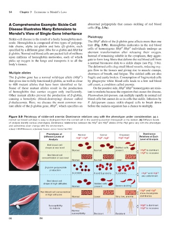
Figure 3.9 Pleiotropy of sickle-cell anemia: Dominance relations vary with the phenotype under consideration. (a) A
normal red blood cell (top) is easy to distinguish from the sickled cell in the scanning electron micrograph at the bottom. (b) Different levels
S
A
of analysis identify various phenotypes. Dominance relationships between the Hbβ and Hbβ alleles of the Hbβ gene vary with the phenotype
and sometimes even change with the environment.
a (top): © BSIP/Newscom; a (bottom): Source: Janice Haney Carr/CDC
Phenotypes at Normal Carrier Diseased Dominance
Di erent Levels of Hb Hb A Hb Hb S Hb Hb S Relations at Each
S
A
A
Analysis Level of Analysis
Red blood cell
shape at sea level Sickled cells
A
Normal Normal present Hb is dominant
Hb is recessive
S
Red blood cell
concentration at sea level
Normal Normal Lower
-globin polypeptide
production
A protein A and S proteins S proteins Hb and Hb S
A
are codominant
Red blood cell
shape at high altitudes Sickled cells
Normal present Severe sickling
A
Hb and Hb
S
Red blood cell concentration
at high altitudes show incomplete
Normal Lower Very low, anemia dominance
S
Susceptibility Hb is dominant
A
to malaria Hb is recessive
Normal
susceptibility Resistant Resistant
(a)
(a) (b)