
Page 19 - Genetics_From_Genes_to_Genomes_6th_FULL_Part1
P. 19
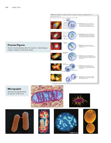
xviii   Guided Tour
                                                             100   Chapter 4  The Chromosome Theory of Inheritance
                                                             Figure 4.10  Mitosis maintains the chromosome number of the parent cell in the two daughter nuclei. In the
                                                             photomicrographs of newt lung cells at the left, chromosomes are stained blue and microtubules appear either green or yellow. Note that the
                                                             drawings are of Ascaris cells (2n = 4).
                                                             a–f: © Photomicrographs by Dr. Conly L. Rieder, Wadsworth Center, Albany, New York 12201-0509
                                                                                 In animal cells
                                                                             Centriole
                                                                              Microtubules
                                                                              Centrosome      (a)   Prophase: (1) Chromosomes condense and
                                                                              Centromere       become visible; (2) centrosomes move apart
                                                                                               toward opposite poles and generate new
                                                                              Chromosome       microtubules; (3) nucleoli begin to disappear.
                                                                             Sister chromatids
                                                                              Nuclear envelope
                                                                             Astral microtubules
                                                                              Kinetochore     (b)   Prometaphase: (1) Nuclear envelope breaks
                                                                                               down; (2) microtubules from the centrosomes
                                                                             Kinetochore       invade the nucleus; (3) sister chromatids attach
                                                                             microtubules      to microtubules from opposite centrosomes.
                                                                              Polar
                                                                              microtubules
                                                                              Metaphase
                                                                              plate
                 Process Figures                                                              (c)   Metaphase: Chromosomes align on the
                                                                                               metaphase plate with sister chromatids facing
                 Step-by-step descriptions allow the student to walk through a                 opposite poles.
                 compact summary of important details.
                                                                             Separating sister
                                                                             chromatids
                                                                                              (d)   Anaphase: (1) The connection between the
                                                                                               centromeres of the sister chromatids is severed;
                                                                                               (2) the now separated sister chromatids move to
                                                                                               opposite poles.

                                                                             Re-forming
                                                                             nuclear envelope
                                                                                              (e)   Telophase: (1) Nuclear membranes and
                                                                                               nucleoli re-form; (2) spindle fibers disappear;
                                                                                               (3) chromosomes uncoil and become a tangle
                                                                                               of chromatin.
                                                                                       Nucleoli reappear
                                                                             Chromatin
                                                                                              (f)    Cytokinesis: The cytoplasm divides, splitting
                                                                                               the elongated parent cell into two daughter
                                                                                               cells with identical nuclei.


                 Micrographs                               har00909_ch04_089-132.indd   100                           12/05/17   6:17 PM

                 Stunning micrographs bring
                 the genetics world to life.