
Page 128 - Genetics_From_Genes_to_Genomes_6th_FULL_Part1
P. 128
120 Chapter 4 The Chromosome Theory of Inheritance
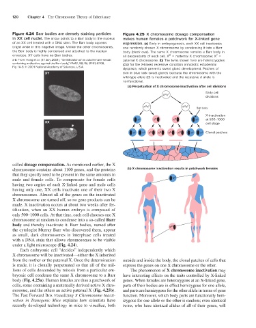
Figure 4.24 Barr bodies are densely staining particles Figure 4.25 X chromosome dosage compensation
in XX cell nuclei. The arrow points to a Barr body in the nucleus makes human females a patchwork for X-linked gene
of an XX cell treated with a DNA stain. The Barr body appears expression. (a) Early in embryogenesis, each XX cell inactivates
bright white in this negative image. Unlike the other chromosomes, one randomly chosen X chromosome by condensing it into a Barr
the Barr body is highly condensed and attached to the nuclear body (black oval). The same X chromosome remains a Barr body in
envelope. XY cells have no Barr bodies. all descendants of each cell. X = maternal X chromosome; X =
M
P
a-b: From: Hong et al. (17 July 2001), “Identification of an autoimmune serum paternal X chromosome. (b) The twins shown here are heterozygotes
containing antibodies against the Barr body,” PNAS, 98(15): 8703-8708, (Dd) for the X-linked recessive condition anhidrotic ectodermal
Fig 1A-B. © 2001 National Academy of Sciences, U.S.A.
dysplasia, which prevents sweat gland development. Patches of
XY skin in blue lack sweat glands because the chromosome with the
wild-type allele (D) is inactivated and the recessive d allele is
nonfunctional.
(a) Perpetuation of X chromosome inactivation after cell divisions
Early cell
M P
X X
divisions
M P
M P
X X X X Barr body
X-inactivation
X p X M X M X p at 500–1000
XX cell stage
X p X p X M X M X M X M X p X p Clonal patches
called dosage compensation. As mentioned earlier, the X
chromosome contains about 1100 genes, and the proteins (b) X chromosome inactivation results in patchwork females
that they specify need to be present in the same amounts in
male and female cells. To compensate for female cells X d
having two copies of each X-linked gene and male cells
having only one, XX cells inactivate one of their two X
chromosomes. Almost all of the genes on the inactivated
X chromosome are turned off, so no gene products can be
made. X inactivation occurs at about two weeks after fer-
tilization, when an XX human embryo is composed of
only 500–1000 cells. At that time, each cell chooses one X
chromosome at random to condense into a so-called Barr
body and thereby inactivate it. Barr bodies, named after X D
the cytologist Murray Barr who discovered them, appear
as small, dark chromosomes in interphase cells treated
with a DNA stain that allows chromosomes to be visible
under a light microscope (Fig. 4.24).
Each embryonic cell “decides” independently which
X chromosome will be inactivated—either the X inherited
from the mother or the paternal X. Once the determination outside and inside the body, the clonal patches of cells that
is made, it is clonally perpetuated so that all of the mil- express the genes on one X chromosome or the other.
lions of cells descended by mitosis from a particular em- The phenomenon of X chromosome inactivation may
bryonic cell condense the same X chromosome to a Barr have interesting effects on the traits controlled by X-linked
body (Fig. 4.25a). Human females are thus a patchwork of genes. When females are heterozygous at an X-linked gene,
cells, some containing a maternally derived active X chro- parts of their bodies are in effect hemizygous for one allele,
mosome, and the others an active paternal X (Fig. 4.25b). and parts are hemizygous for the other allele in terms of gene
The Fast Forward Box Visualizing X Chromosome Inacti- function. Moreover, which body parts are functionally hem-
vation in Transgenic Mice explains how scientists have izygous for one allele or the other is random; even identical
recently developed technology in mice to visualize, both twins, who have identical alleles of all of their genes, will