
Page 37 - Genetics_From_Genes_to_Genomes_6th_FULL_Part2
P. 37
196 Chapter 6 DNA Structure, Replication, and Recombination
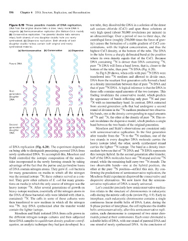
Figure 6.19 Three possible models of DNA replication. test tube, they dissolved the DNA in a solution of the dense
DNA from the original double helix is blue; newly made DNA is salt cesium chloride (CsCl) and spun these solutions at
magenta. (a) Semiconservative replication (the Watson-Crick model). very high speed (about 50,000 revolutions per minute) in
(b) Conservative replication: The parental double helix remains an ultracentrifuge. Over a period of two to three days, the
intact; both strands of one daughter double helix are newly
synthesized. (c) Dispersive replication: Both strands of both centrifugal force (roughly 250,000 times the force of grav-
daughter double helixes contain both original and newly ity) causes the formation of a stable gradient of CsCl con-
synthesized material. centrations, with the highest concentration, and thus the
(a) Semiconservative (b) Conservative (c) Dispersive highest CsCl density, at the bottom of the tube. The DNA
in the tube forms a sharply delineated band at the position
where its own density equals that of the CsCl. Because
15
14
DNA containing N is denser than DNA containing N,
Parent DNA
15
pure N DNA will form a band lower, that is, closer to the
14
bottom of the tube, than pure N DNA (Fig. 6.20).
15
As Fig 6.20 shows, when cells with pure N DNA were
14
transferred into N medium and allowed to divide once,
DNA from the resultant first-generation cells formed a band
15
at a density intermediate between that of pure N DNA and
14
that of pure N DNA. A logical inference is that the DNA in
First- these cells contains equal amounts of the two isotopes. This
generation finding invalidates the conservative model, which predicts
daughter 14
DNA the appearance of bands reflecting only pure N and pure
15 N with no intermediary band. In contrast, DNA extracted
from second-generation cells that had undergone a second
14
round of division in the N medium produced two observa-
ble bands, one at the density corresponding to equal amounts
14
14
15
of N and N, the other at the density of pure N. This re-
sult invalidates the dispersive model, which predicts a single
Second- band between the two bands of the original generation.
generation Meselson and Stahl’s observations are consistent only
daughter
DNA with semiconservative replication. In the first generation
14
15
after transfer from the N to the N medium, one of the
two strands in every daughter DNA molecule carries the
heavy isotope label; the other, newly synthesized strand
14
of DNA replication (Fig. 6.20). The experiment depended carries the lighter N isotope. The band at a density inter-
15
14
on being able to distinguish preexisting parental DNA from mediate between that of N DNA and N DNA represents
newly synthesized DNA. To accomplish this, Meselson and this isotopic hybrid. In the second generation after transfer,
14
15
Stahl controlled the isotopic composition of the nucleo- half of the DNA molecules have one N strand and one N
14
tides incorporated in the newly forming strands by taking strand, while the remaining half carry two N strands. The
advantage of the fact that the purine and pyrimidine bases two observable bands—one at the hybrid position, the
14
of DNA contain nitrogen atoms. They grew E. coli bacteria other at the pure N position—reflect this mix. By con-
for many generations on media in which all the nitrogen firming the predictions of semiconservative replication, the
14
was the normal isotope N; these cultures served as a con- Meselson-Stahl experiment disproved the conservative and
trol. They grew other cultures of E. coli for many genera- dispersive alternatives. We now know that the semicon-
tions on media in which the only source of nitrogen was the servative replication of DNA is nearly universal.
15
heavy isotope N. After several generations of growth on Let’s consider precisely how semiconservative replica-
heavy-isotope medium, essentially all the nitrogen atoms in tion relates to the structure of chromosomes in eukaryotic
the DNA of these bacterial cells were labeled with (that is, cells during the mitotic cell cycle (review Fig. 4.9). Early in
15
contained) N. The cells in some of these cultures were interphase, each eukaryotic chromosome contains a single
then transferred to new medium in which all the nitrogen continuous linear double helix of DNA. Later, during the
14
was N. Any DNA synthesized after the transfer would S-phase portion of interphase, the cell replicates the double
contain the lighter isotope. helix semiconservatively; after this semiconservative repli-
Meselson and Stahl isolated DNA from cells grown in cation, each chromosome is composed of two sister chro-
the different nitrogen-isotope cultures and then subjected matids joined at their centromeres. Each sister chromatid is
these DNA samples to equilibrium density gradient centrif- a double helix of DNA, with one strand of parental DNA and
ugation, an analytic technique they had just developed. In a one strand of newly synthesized DNA. At the conclusion of