
Page 84 - Genetics_From_Genes_to_Genomes_6th_FULL_Part2
P. 84
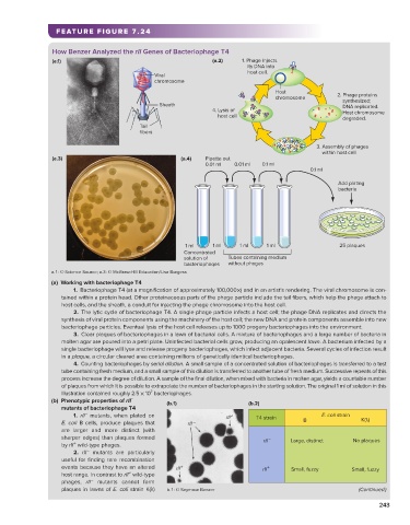
FEATURE FIGURE 7.24

                        How Benzer Analyzed the rII Genes of Bacteriophage T4
                        (a.1)                                           (a.2)    1. Phage injects
                                                                                     its DNA into
                                                                                     host cell.
                                                      Viral
                                                      chromosome
                                                                                          Host
                                                                                          chromosome         2. Phage proteins
                                                                                                                 synthesized;
                                                        Sheath                                                   DNA replicated.
                                                                        4. Lysis of                              Host chromosome
                                                                            host cell
                                                                                                                 degraded.
                                                  Tail
                                                  fibers
                                                                                                       3. Assembly of phages
                                                                                                           within host cell
                        (a.3)                                 (a.4)   Pipette out
                                                                      0.01 ml  0.01 ml  0.1 ml
                                                                                                     0.1 ml
                                                                                                             Add plating
                                                                                                             bacteria


                                                                1 ml    1 ml    1 ml    1 ml                  25 plaques
                                                               Concentrated
                                                               solution of   Tubes containing medium
                                                               bacteriophages  without phages
                        a.1: © Science Source; a.3: © McGraw-Hill Education/Lisa Burgess
                        (a)  Working with bacteriophage T4
                              1.  Bacteriophage T4 (at a magnification of approximately 100,000×) and in an artist’s rendering. The viral chromosome is con-
                           tained within a protein head. Other proteinaceous parts of the phage particle include the tail fibers, which help the phage attach to
                           host cells, and the sheath, a conduit for injecting the phage chromosome into the host cell.
                              2.  The lytic cycle of bacteriophage T4. A single phage particle infects a host cell; the phage DNA replicates and directs the
                           synthesis of viral protein components using the machinery of the host cell; the new DNA and protein components assemble into new
                           bacteriophage particles. Eventual lysis of the host cell releases up to 1000 progeny bacteriophages into the environment.
                              3.  Clear plaques of bacteriophages in a lawn of bacterial cells. A mixture of bacteriophages and a large number of bacteria in
                           molten agar are poured into a petri plate. Uninfected bacterial cells grow, producing an opalescent lawn. A bacterium infected by a
                           single bacteriophage will lyse and release progeny bacteriophages, which infect adjacent bacteria. Several cycles of infection result
                           in a plaque, a circular cleared area containing millions of genetically identical bacteriophages.
                              4.  Counting bacteriophages by serial dilution. A small sample of a concentrated solution of bacteriophages is transferred to a test
                           tube containing fresh medium, and a small sample of this dilution is transferred to another tube of fresh medium. Successive repeats of this
                           process increase the degree of dilution. A sample of the final dilution, when mixed with bacteria in molten agar, yields a countable number
                           of plaques from which it is possible to extrapolate the number of bacteriophages in the starting solution. The original 1 ml of solution in this
                                                     7
                             illustration contained roughly 2.5 × 10  bacteriophages.
                                               −
                        (b)   Phenotypic properties of rII     (b.1)              (b.2)
                          mutants of bacteriophage T4
                                  −
                              1.  rII  mutants, when plated on              rII +    T4 strain           E. coli strain
                           E. coli B cells, produce plaques that   rII –                           B                K( )
                           are larger and more distinct (with
                           sharper edges) than plaques formed                           –      Large, distinct    No plaques
                              +
                           by rII  wild-type phages.                                   rII
                                  −
                              2.  rII  mutants are particularly
                           useful for finding rare recombination
                           events because they have an altered   rII +                 rII +   Small, fuzzy      Small, fuzzy
                                               +
                           host range. In contrast to rII  wild-type
                                   −
                           phages,  rII  mutants cannot form
                           plaques in lawns of E. coli strain K(λ)   b.1: © Seymour Benzer                         (Continued)
                                                                                                                         243