
Page 188 - Genetics_From_Genes_to_Genomes_6th_FULL_Part1
P. 188
180 Chapter 5 Linkage, Recombination, and the Mapping of Genes on Chromosomes
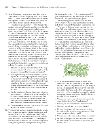
51. Neurofibromas are tumors of the skin that can arise like Drosophila or mice. Cells expressing this GFP
−
+
when a skin cell that is originally NF1 / NF1 loses gene will glow green in the microscope, while those
+
the NF1 allele. This wild-type allele encodes a func- without the GFP gene will not glow green.
tional protein (called a tumor suppressor), while the Mice homozygous for the recessive mutation
−
NF1 allele encodes a nonfunctional protein. small cells (smc) die as early embryos because their
−
+
A patient of genotype NF1 / NF1 has 20 inde- cells divide prematurely before they reach normal size.
pendent tumors in different areas of the skin. Samples You want to design a mouse carrying one copy
are taken of normal, noncancerous cells from this of the GFP gene and heterozygous for smc in which
patient, as well as of cells from each of the 20 tumors. you could generate clones in adult mice by mitotic
Extracts of these samples are analyzed by a technique recombination. In this designer mouse, every cell in
called gel electrophoresis that can detect variant every clone that is not green would be homozygous
forms of four different proteins (A, B, C, and D) all for the smc mutation. The figure below shows a field
encoded by genes that lie on the same autosome as of epithelial cells in the mouse you design. You will
NF1. Each protein has a slow (S) and a fast (F) form see some cells that are normal size and other cells
that are encoded by different alleles (for example, A that are small. You will also see cells of three differ-
S
and A ). In the extract of normal tissue, slow and fast ent colors: blank, weakly glowing cells (light green),
F
variants of all four proteins are found. In the extracts and brightly glowing cells (dark green). Most of the
of the tumors, 12 had only the fast variants of proteins cells in the epithelium of this mouse are of normal
A and D but both the fast and slow variants of pro- size and weakly glowing. The epithelium also
teins B and C; 6 had only the fast variant of protein A contains three clones of cells (1, 2, and 3) that have
but both the fast and slow variants of proteins B, C, unusual appearances due to the occurrence of
and D; and the remaining 2 tumor extracts had only mitotic recombination.
the fast variant of protein A, only the slow variant of
protein B, the fast and slow variants of protein C, and
only the fast variant of protein D. 1
a. What kind of genetic event described in this chap- 3
ter could cause all 20 tumors, assuming that all the
tumors are produced by the same mechanism?
b. Draw a genetic map describing these data, assum-
ing that this small sample represents all the types 2
of tumors that could be formed by the same mech-
anism in this patient. Show which alleles of which a. Show the chromosomes and centromeres, the
genes lie on the two homologous chromosomes. alleles smc and smc, and GFP (GFP gene pres-
+
+
Indicate all relative distances that can be estimated. ent) and GFP (GFP gene absent) in your designer
−
Note that NF1 is one of the genes you can map in mouse. (As a reminder, this mouse will carry one
this way. copy of the GFP gene and will be heterozygous for
c. Another mechanism that can lead to neurofibromas smc. Every cell in every clone generated by mitotic
in this patient is a mitotic error producing cells with recombination that is not green should be homozy-
45 rather than the normal 46 chromosomes. How gous for the smc mutation.)
can this mechanism cause tumors? How do you b. Why do you need to use mitotic recombination to
know, just from the results described, that none of study the function of smc in adult mice?
+
these 20 tumors is formed by such mitotic errors? c. Why do you see cells of three different colors?
d. Can you think of any other type of error that could d. Why are clones 1 and 2 next to each other?
produce the results described?
52. Two important methods for understanding the genetic e. On your map in part (a), place an arrow to show
the position of a mitotic recombination event that
basis for development are mitotic crossing-over and could give rise to clones 1 and 2.
the use of the gene from jellyfish called GFP (for
green fluorescent protein) that makes these animals f. Why do more cells exist in clone 1 than in clone 2?
glow in the dark. By recombinant DNA techniques g. On your map in part (a), place an arrow to show
described later in the book, you can insert the jellyfish the position of a mitotic crossover that could give
GFP gene anywhere into the genome of organisms rise to clone 3.